An Anatomical Atlas of the 18th Century
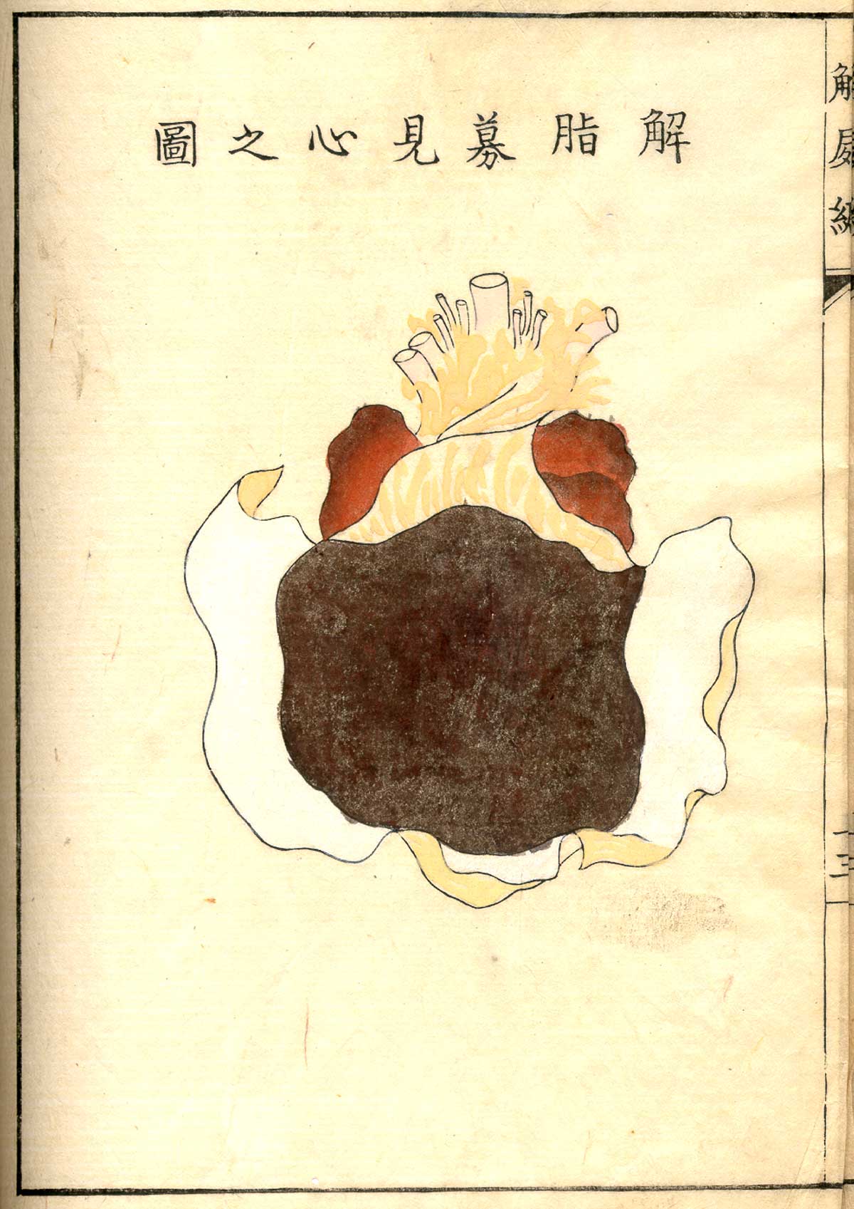
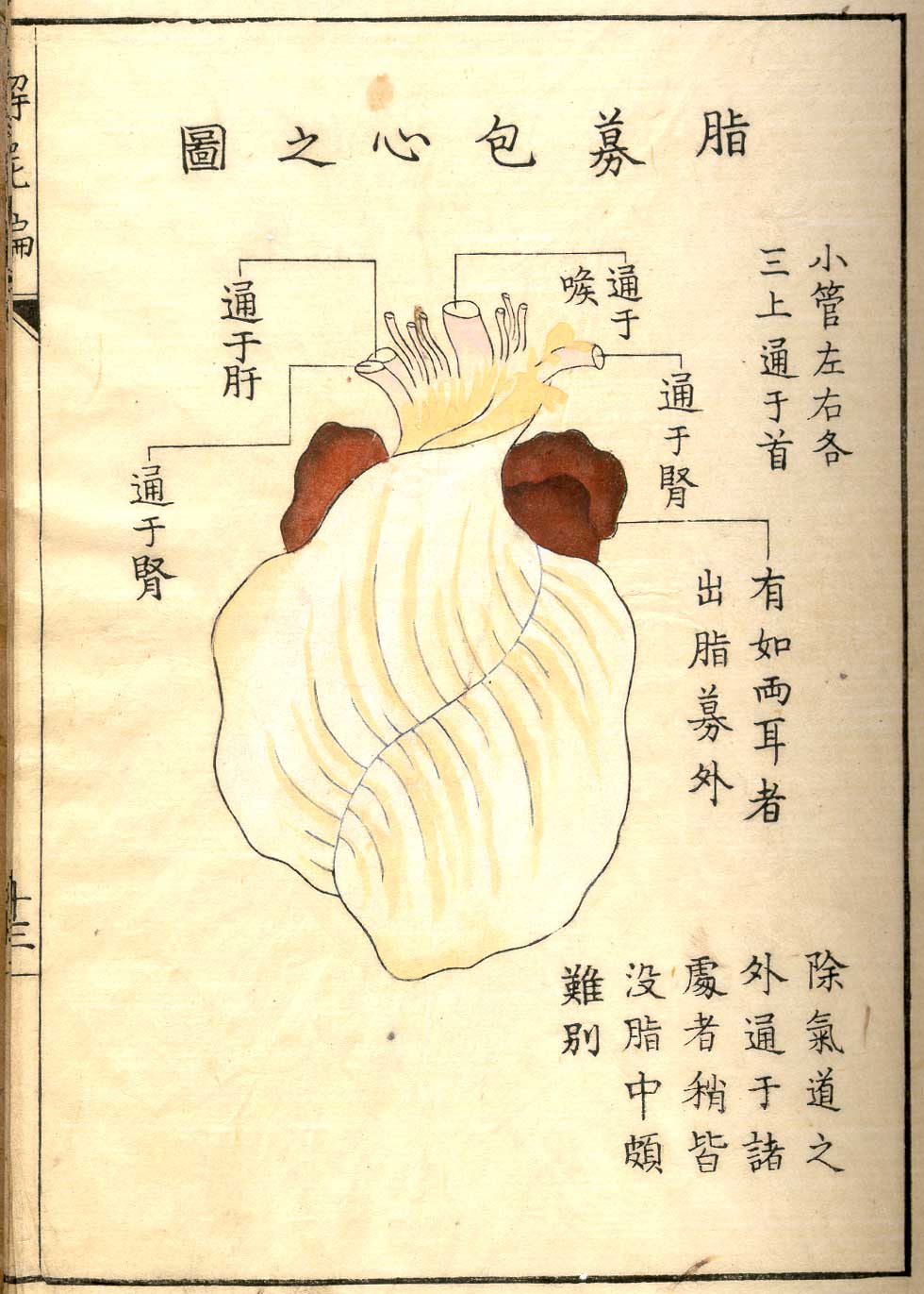
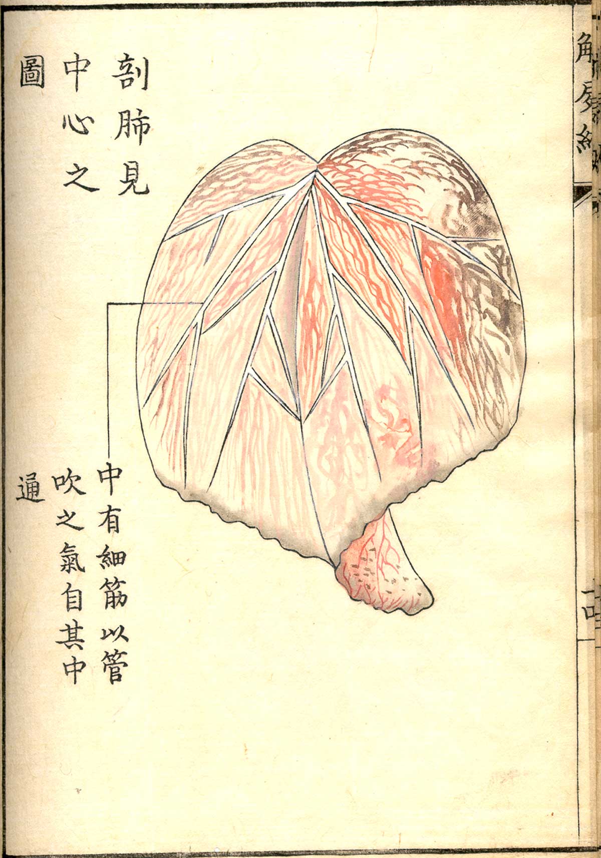
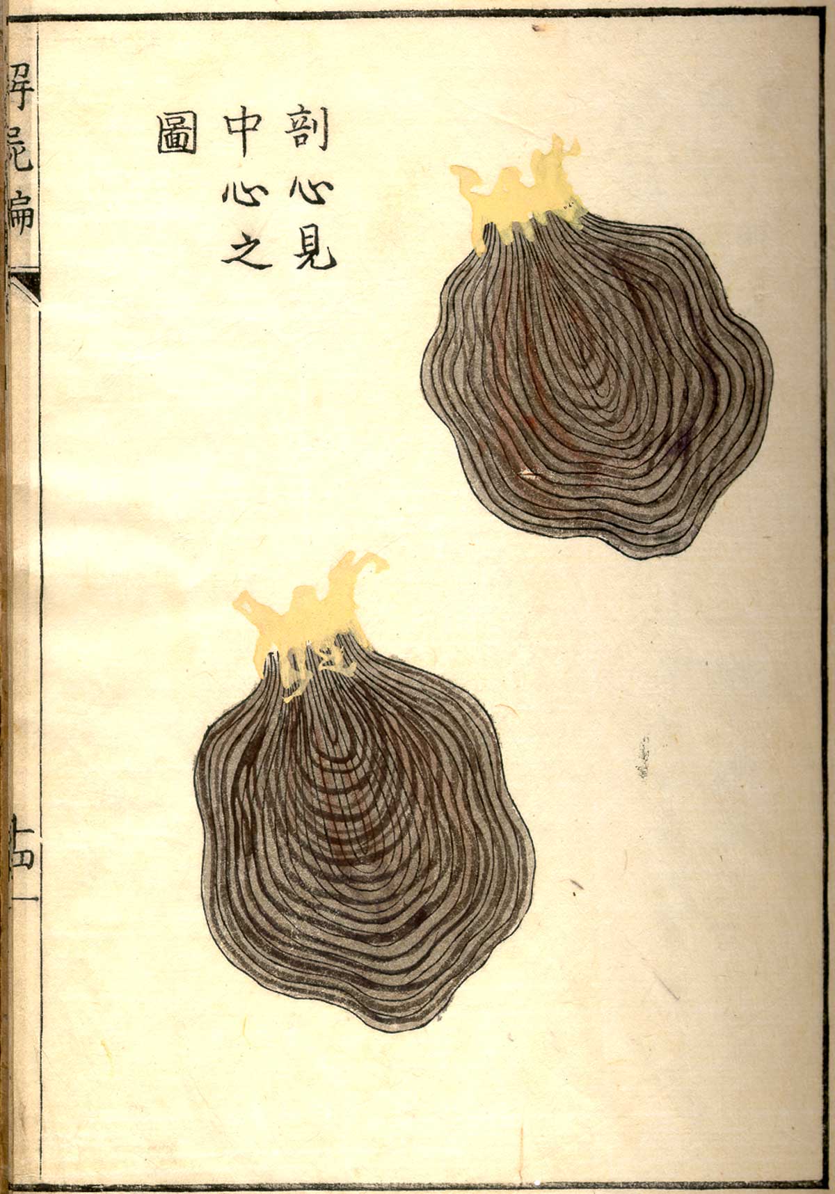
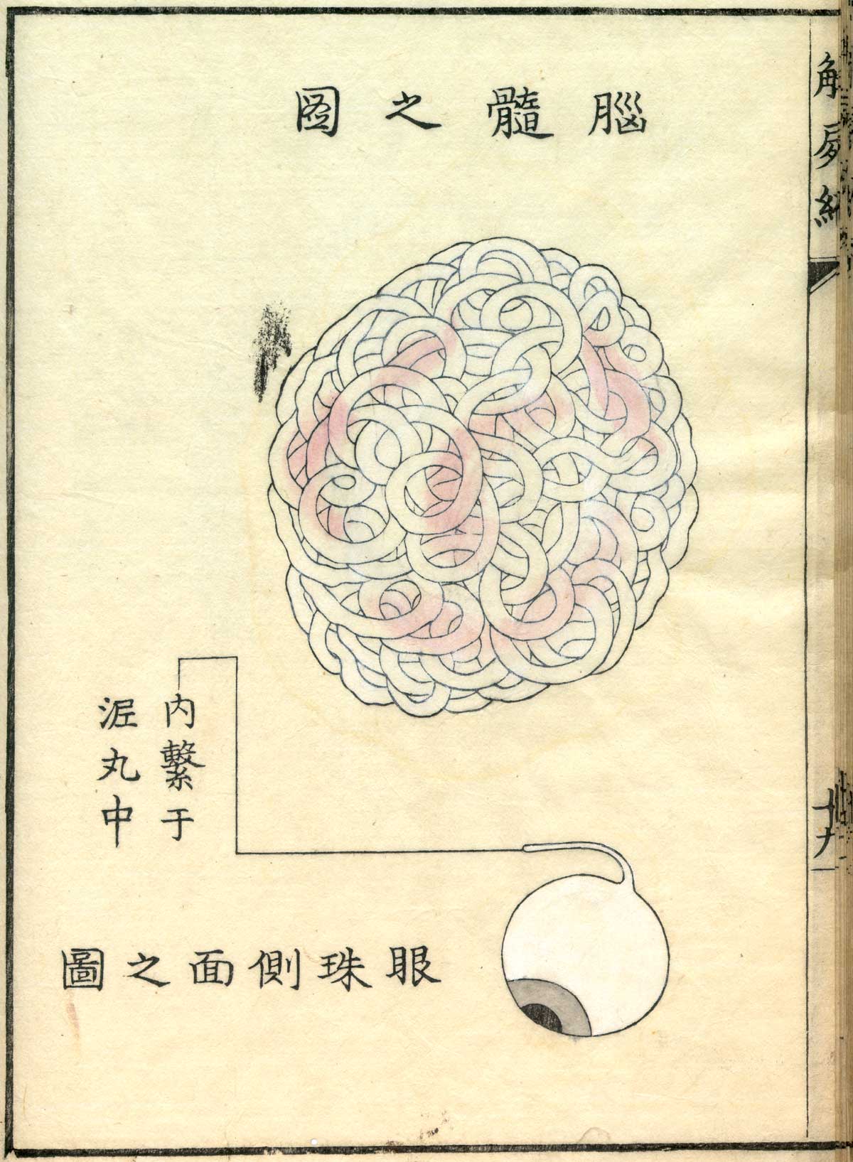
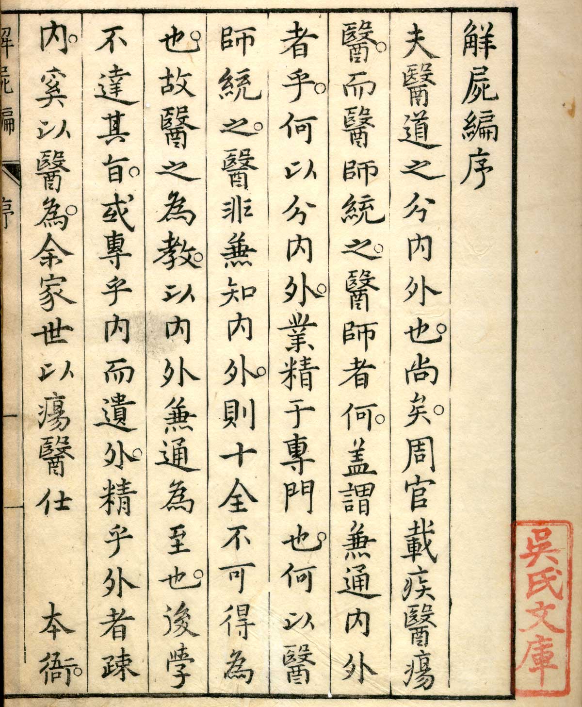
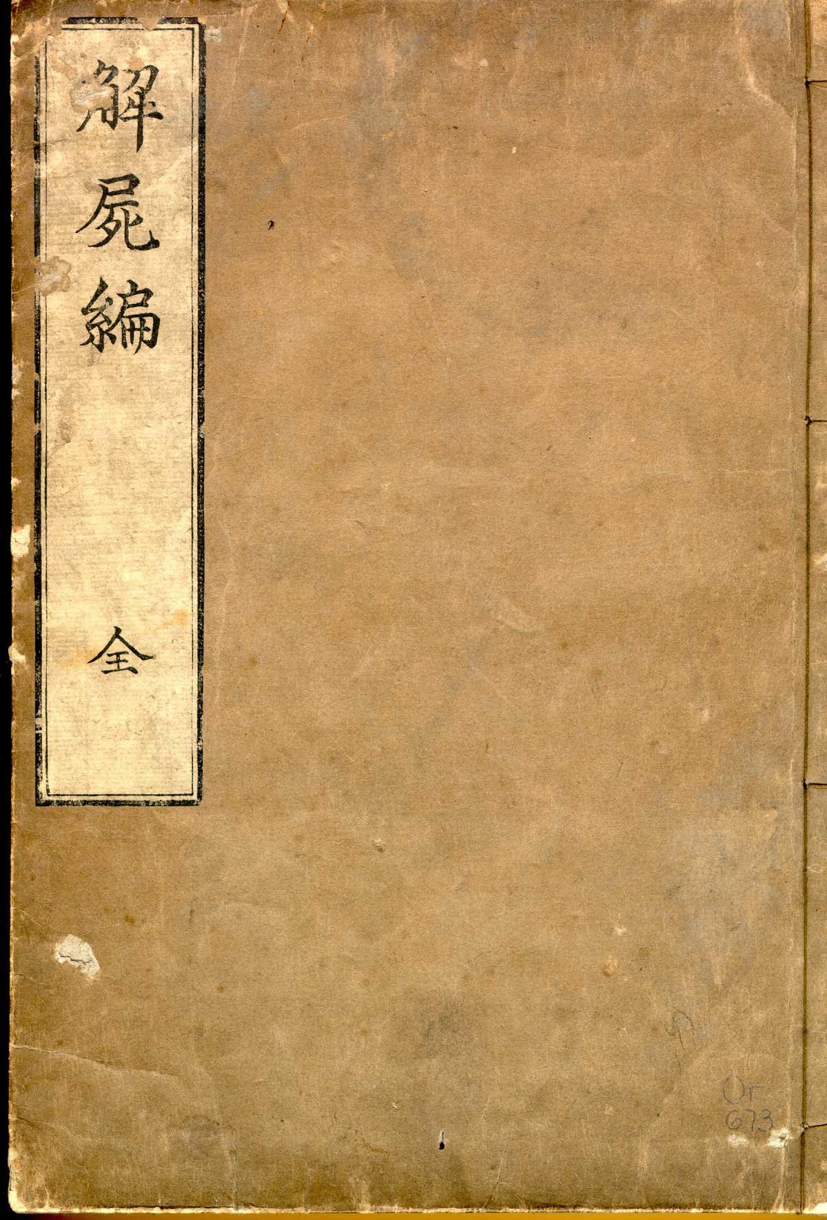
The illustrations engraved on wood in 'Kaishi Hen' are the vestiges of the first human dissections that took place in Japan.
© Public Domain
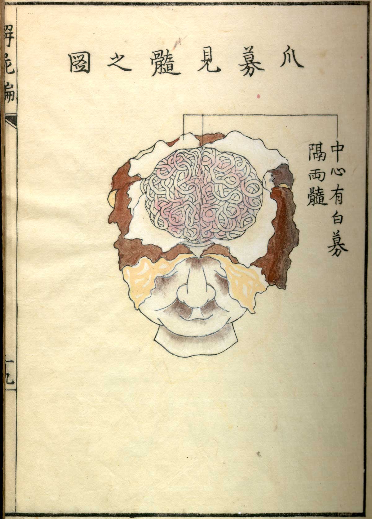
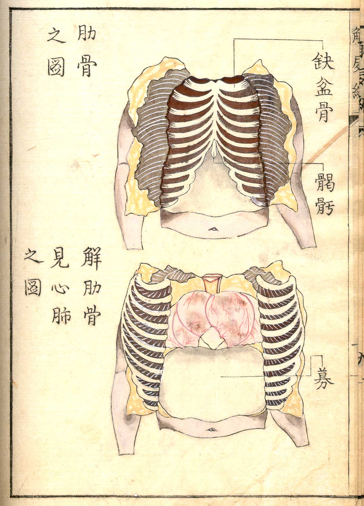
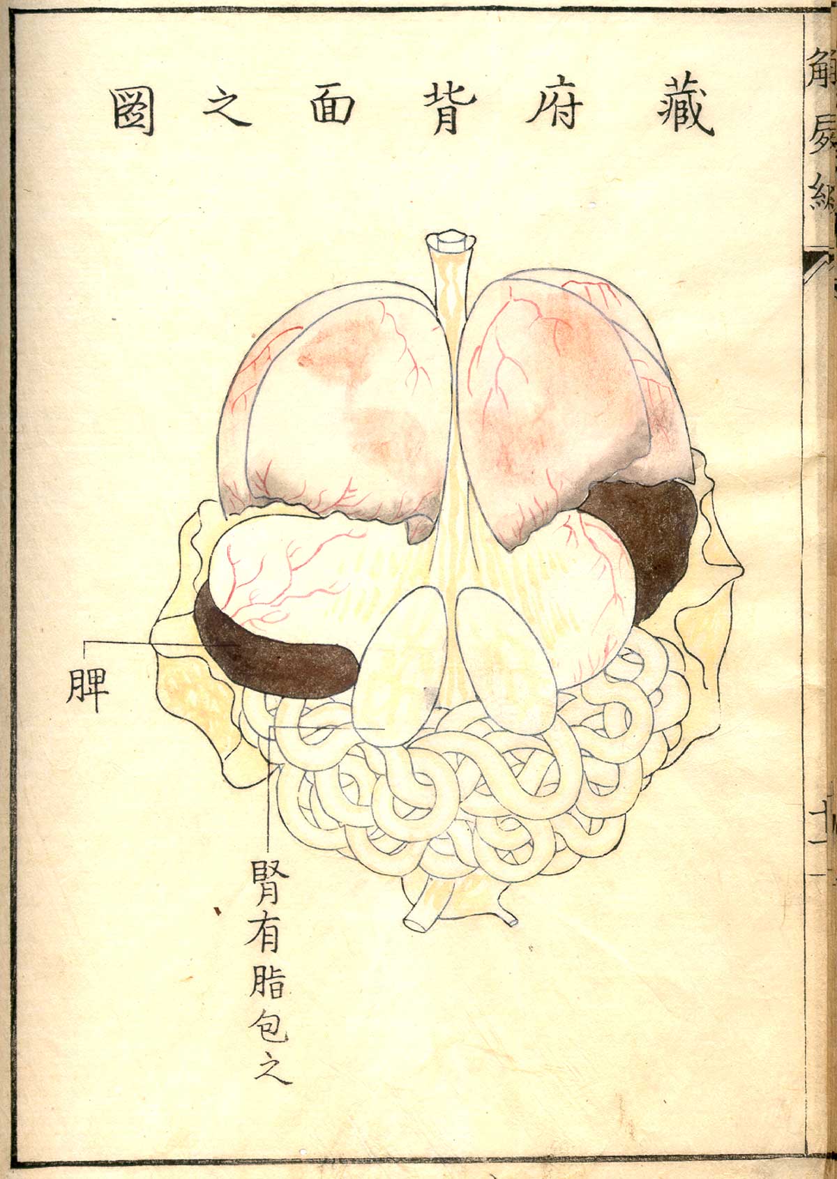
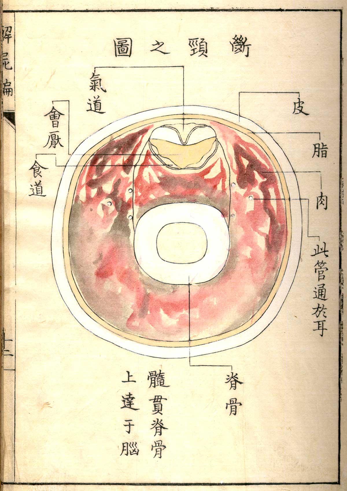
This series of 23 illustrations, compiled in an anatomical atlas entitled Kaishi Hen, represents various cross-sections of the human body. These hand-coloured wood engravings are the work of Japanese painter and sculptor Aoki Shukuya, and are based on sketches by Shinnin Kawaguchi, a disciple of Gengai Ogino, a Japanese doctor who studied Western medical theory.
The beginnings of experimental medicine
This anatomical atlas, published in 1772, marked the beginnings of experimental medicine in Japan. Its illustrations of intestines, the brain, the skeleton, and lungs depict the dissection of the body of a criminal executed in Kyoto two years earlier. At the time, this was only the third human dissection to have been carried out in Japan, the first having taken place in 1750 and also having been documented through wood engravings, carried out by artist Asanuma Suketsune and included in the book A Galaxy of Old Japanese Medical Books I. Dissection had been forbidden in the country since antiquity, and was only officially authorised in 1858.
Kaishi Hen (1772) is an anatomical atlas engraved on wood by Aoki Shukuya and digitised by the United States National Library of Medicine.
© Public Domain
© Public Domain
© Public Domain
© Public Domain
© Public Domain
© Public Domain
© Public Domain
© Public Domain
© Public Domain
© Public Domain